São Paulo-Würzburg Atlas of Neuroanatomy
SWAN
A multimodal human neuroanatomy atlas project linking high-resolution histology with blockface imaging, in situ specimen MRI, DTI, and MNI space.
Overview
A bridge between laboratory neuroanatomy and imaging space
SWAN is designed to connect microscopic human neuroanatomy with modern brain imaging in a documented, registration-aware framework.
We bring together high-thickness histological sections imaged with brightfield microscopy, darkfield microscopy of selected subcortical and brainstem regions, blockface imaging, in situ specimen MRI, DTI, and MNI-space registration for anatomical interpretation and future open-science release.
Microscopic anatomy in imaging space
SWAN connects tissue-level anatomical detail with specimen-specific MRI and standardized stereotactic coordinates.
Multimodal correspondence
Brightfield histology, darkfield microscopy, blockface imaging, in situ specimen MRI, DTI, and MNI space are treated as linked but distinct sources of anatomical information.
Registration-aware atlas building
We explicitly address two-dimensional section deformation and three-dimensional specimen-to-MRI alignment.
Open-science trajectory
We are preparing the dataset and documentation for staged public release after review and publication.
Multimodal pipeline
From microscopy to standardized neuroimaging coordinates
- 01
Brightfield histology and darkfield microscopy
Microscopic and fiber-rich anatomical contrast from human brain sections.
- 02
Blockface imaging
An intermediate reference for preserving section geometry during tissue processing.
- 03
In situ specimen MRI and DTI
Specimen-specific volumetric imaging that bridges processed tissue and neuroimaging space.
- 04
MNI-space registration
A standardized coordinate framework for representative anatomical comparison.
Brightfield histology / darkfield microscopy -> blockface -> in situ specimen MRI / DTI -> MNI space
Representative images
Curated first-version visual material
We emphasize representative media: microscopy, registration, MNI-space correspondence, and quality-control examples.
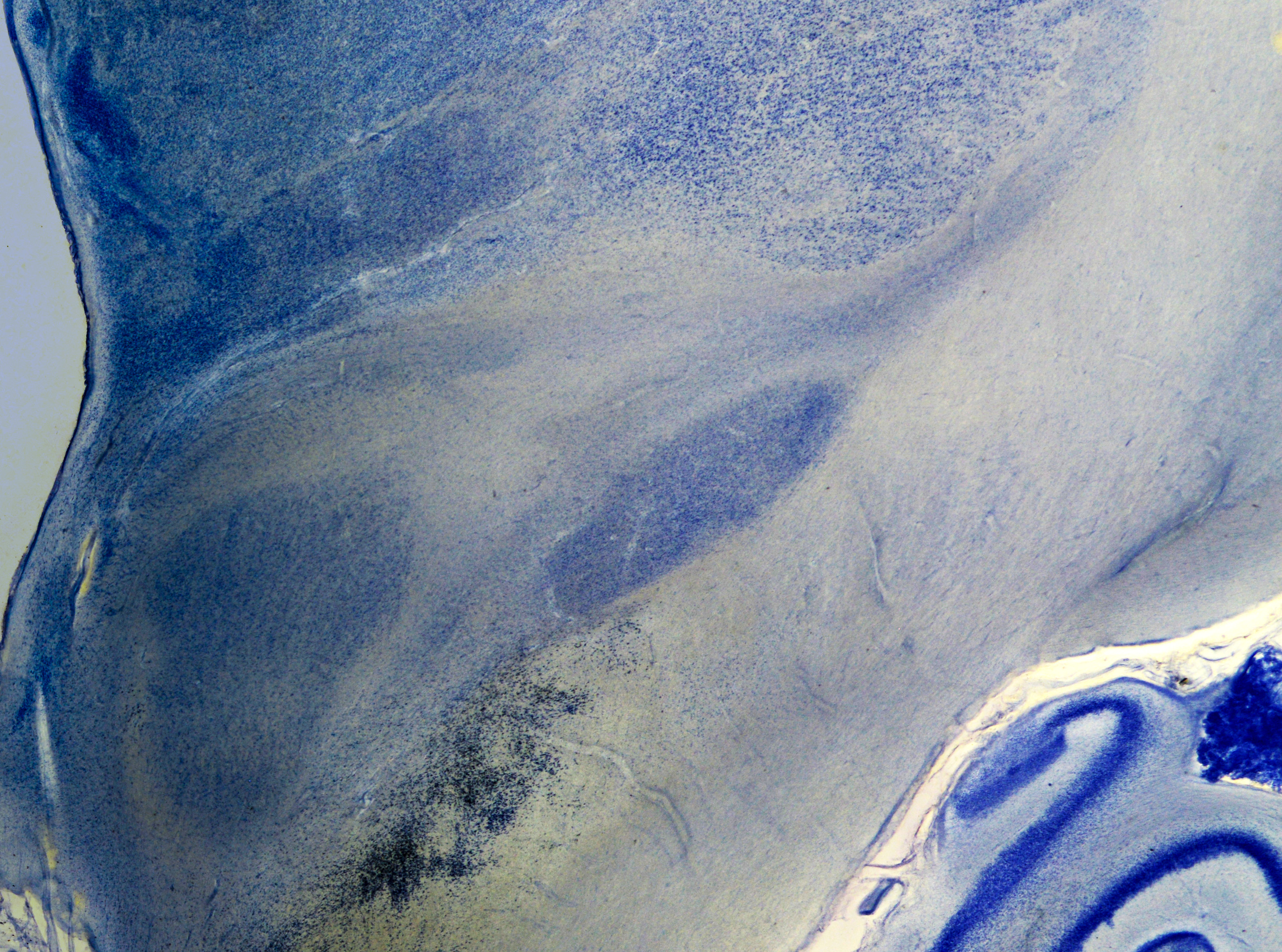
High-resolution histological contrast supporting anatomical annotation and multimodal registration.
Complementary contrast for selected subcortical, diencephalic, and brainstem anatomy.
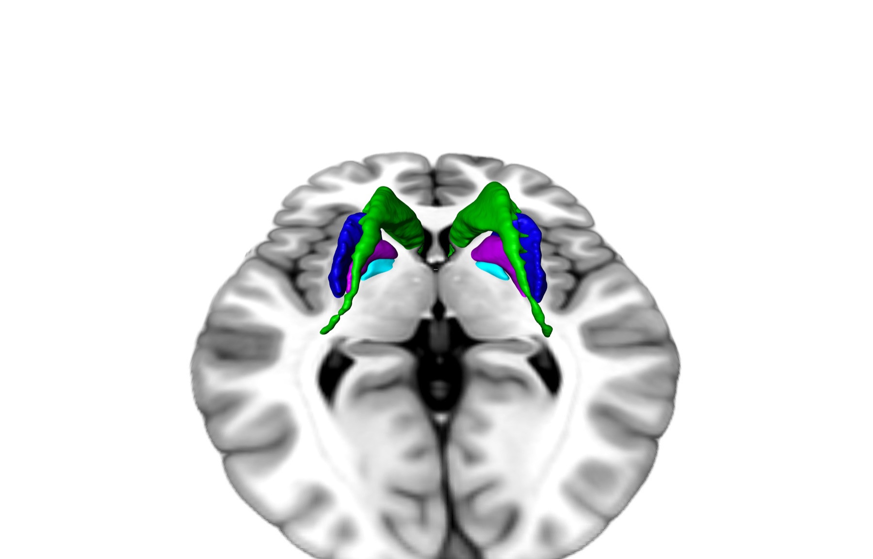
Representative standard-space registration for neuroimaging interoperability.
SWAN Viewer