
Project
Origin and motivation
SWAN began from a practical problem in functional neurosurgery: structures that matter for stereotactic targeting can be small, complex, variable, and difficult to resolve completely with conventional imaging or classical atlas registration. We developed SWAN to create a more spatially coherent bridge between histology and imaging when individual anatomy and atlas-based expectations do not fully agree.
The central scientific challenge is not simply to make another atlas image set. It is to preserve the relationship between microscopic anatomy, tissue-processing deformation, the original specimen geometry, in situ specimen MRI, and standardized neuroimaging space.
Why SWAN was needed
Classical stereotactic atlases have been foundational for functional neurosurgery, but printed histological plates and digitized atlas reconstructions may be limited by sparse sampling, tissue deformation, and imperfect three-dimensional consistency. MRI-based atlases provide essential in vivo anatomical context, but MRI and histology have different physical origins and reveal different aspects of tissue organization.
We created SWAN to address this gap by linking high-resolution brightfield histological information to blockface images, in situ specimen MRI, DTI, and MNI space through a documented multimodal registration pipeline.
São Paulo–Würzburg collaboration
The project grew from a long-standing collaboration between São Paulo and Würzburg, combining functional neurosurgery, neuropathology, brain banking, high-thickness histological processing, radiology, image registration, and computational neuroanatomy. Our collaboration brings together contributors associated with the University of São Paulo, the University of Würzburg, InRad/HCFMUSP, Mevis Informática Médica, Lead-DBS and Network Stimulation collaborators, UCSF, Charité - Universitätsmedizin Berlin, Brigham and Women’s Hospital / Harvard Medical School, and University Hospital Cologne.
SWAN is presented as a multidisciplinary scientific project.
Laboratory neuroanatomy and clinical imaging
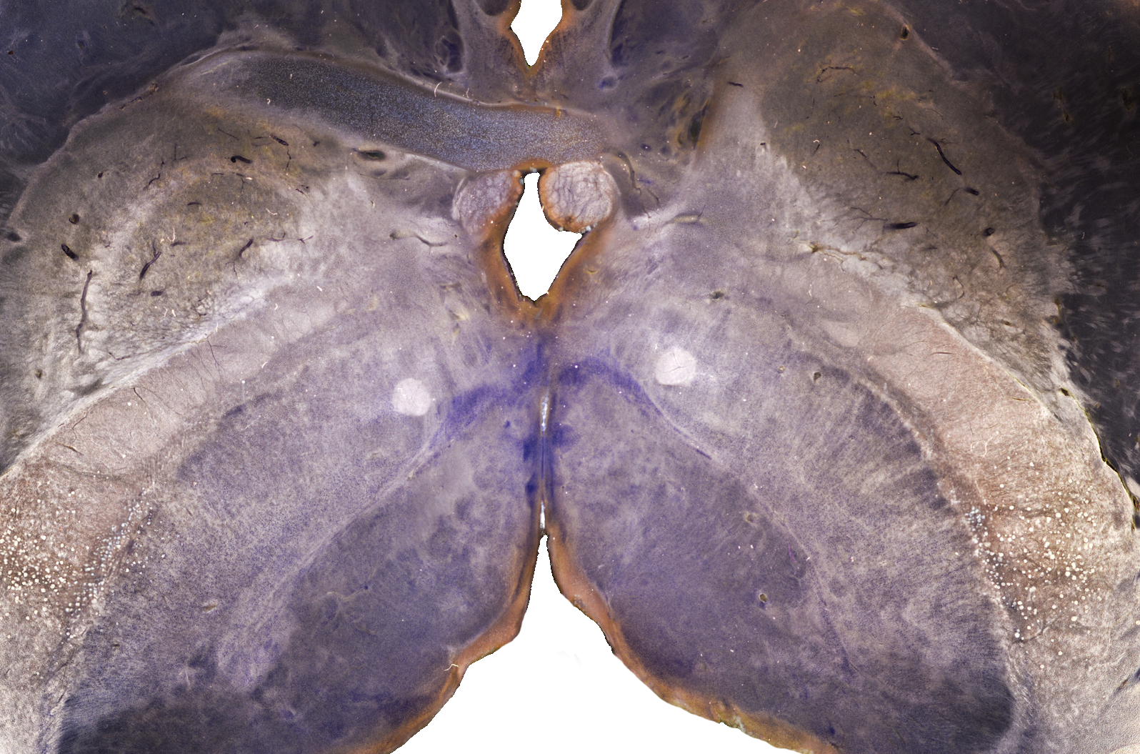
SWAN is positioned at the interface between laboratory neuroanatomy and clinical/research imaging. High-resolution brightfield histological sections provide microscopic anatomical detail. Darkfield microscopy provides complementary contrast for selected fiber-rich subcortical and brainstem regions. Blockface imaging helps preserve the geometry of the specimen during sectioning. In situ specimen MRI and DTI connect the same specimen to volumetric imaging. MNI-space registration provides a bridge to standardized neuroimaging coordinates.
This relationship is especially relevant for research in functional neurosurgery and deep brain stimulation, where microscopic anatomy, electrode localization, stimulation fields, and patient imaging must be interpreted across different spatial scales.
