
Gallery
Representative images from the SWAN project
A curated visual overview of first-version media, emphasizing microscopy, registration, MNI-space correspondence, and quality control.
Curated first release
Images for scientific context
The first SWAN gallery is not an exhaustive atlas release. It presents selected representative material suitable for a reviewer-facing public website.
Brightfield Histology
Microscopic anatomical contrast motivating the SWAN atlas framework.
Brightfield histology showing tissue-level detail relevant for annotation and multimodal registration.
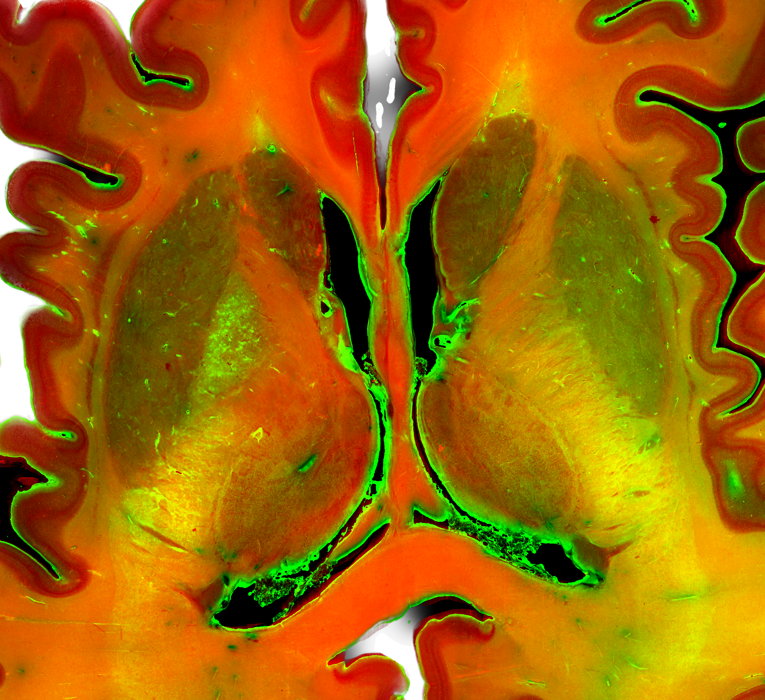
Darkfield Microscopy
Complementary contrast for selected subcortical, diencephalic, and brainstem anatomy.
Darkfield microscopy contributes additional anatomical contrast for interpretation and registration.
Blockface Imaging
Intermediate anatomical reference between gross tissue geometry and processed histological sections.
A visual teaser for orthogonal navigation concepts in the SWAN Viewer pipeline.
Registration
Example of slice-wise alignment supporting local deformation correction.
Correspondence between reconstructed histological information and specimen-specific imaging space.
Coordinate-space bridge between microscopic anatomy and standardized neuroimaging.
MNI Space and Quality Control
Conceptual standard-space interoperability without implying that all public downloads are already available.
Reviewer-facing transparency about registration performance, not clinical validation.
SWAN Viewer Teaser
A preview of modality switching concepts for the SWAN Viewer.
Viewer teaser for reconstructed histological navigation and modality comparison.
Secondary teaser for standard-space visualization in the SWAN Viewer pipeline.
