
Methods
Overview
We designed the SWAN pipeline to link microscopic neuroanatomy with volumetric imaging and standardized stereotactic space. The workflow preserves the distinction between tissue-derived image modalities, specimen imaging, in situ specimen MRI, and MNI-space registration.
The high-level registration chain is:
Brightfield histology / darkfield microscopy -> blockface -> in situ specimen MRI / DTI -> MNI space
Modalities
Histology
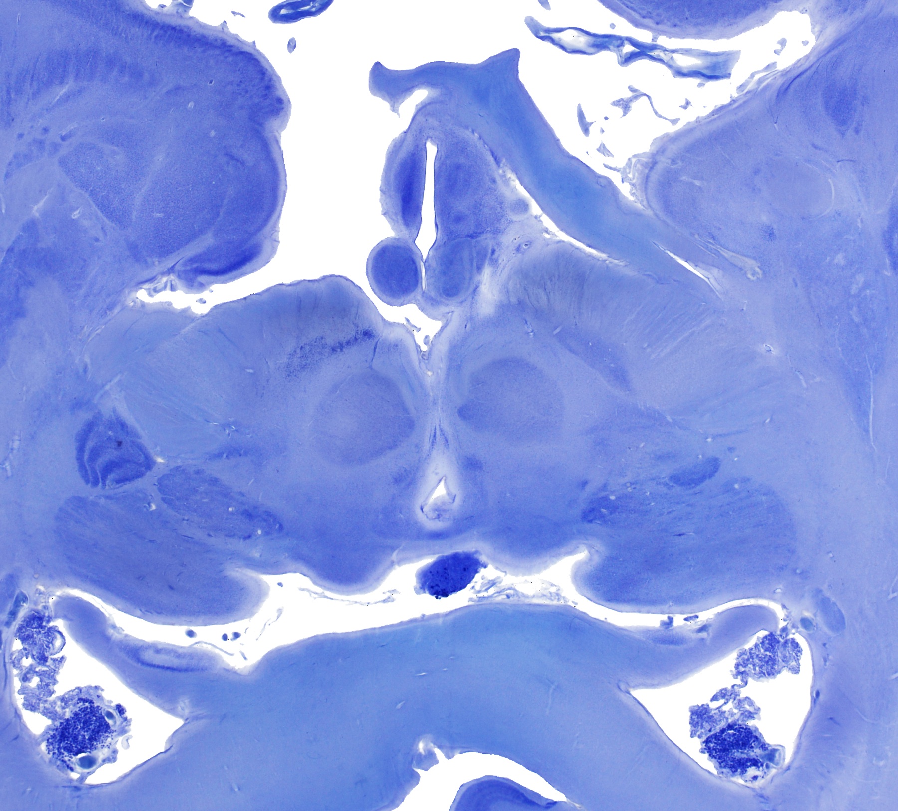
Our histological dataset includes high-thickness human brain sections stained for cellular and cytoarchitectural contrast and imaged with brightfield microscopy. The current dataset includes 430 micrometer sections and gallocyanin / modified Nissl staining.
Darkfield microscopy
Darkfield microscopy provides complementary contrast for selected subcortical, diencephalic, and brainstem regions. It is especially useful for highlighting fiber-rich anatomy and boundaries that may be less conspicuous in a single optical modality. Darkfield coverage is described conservatively as selected regional coverage.
Blockface imaging
Blockface images are acquired during tissue processing before histological staining and mounting. They provide an intermediate anatomical reference for section geometry and help bridge local histological deformation with the three-dimensional specimen.
In situ specimen MRI and DTI
The in situ specimen MRI is described as a 3T MRI of the same brain before extraction from the skull. This imaging space is used as a specimen-specific bridge between processed histological material and volumetric neuroimaging. DTI is part of the multimodal framework.
MNI space
MNI space provides a standardized reference for comparing SWAN-derived anatomy with neuroimaging datasets and individual imaging. Representative MNI-space labels and segmentations are part of the SWAN framework, with public release details to be finalized through the planned repository.
Pipeline explanation
We address deformation at multiple stages. Two-dimensional registration focuses on slice-wise alignment of brightfield histology and darkfield microscopy images to blockface reference images. Three-dimensional registration links the reconstructed histological/blockface dataset to in situ specimen MRI. MNI-space registration then supports standardized anatomical comparison.
Future releases aim to include effective forward and inverse composite transforms linking histology/SWAN and MNI space.
2D slice-wise registration
The 2D registration step aligns individual brightfield histological and darkfield microscopy sections with their corresponding blockface images. This step is intended to correct local deformation introduced by cutting, staining, mounting, coverslipping, and image digitization. The workflow includes computational registration, expert review, and manual corrections where needed.
We include 2D slice-wise registration and quality-control outputs as core parts of the SWAN pipeline.
3D registration
The 3D registration step reconstructs registered sections into a volumetric dataset and aligns the histology/blockface volume with the in situ specimen MRI. This step addresses deformation related to extraction, fixation, dehydration, embedding, and blocking. It provides the bridge from processed tissue to specimen-specific MRI space.
MNI-space registration
MNI-space registration links the reconstructed SWAN dataset to standardized stereotactic coordinates. This enables representative histology-derived labels and segmentations to be interpreted alongside neuroimaging data. The exact public release scope of repository files remains to be finalized.
Quality control
Quality control combines visual review and quantitative assessment. Representative outputs include:
- Histology-to-blockface overlays
- Darkfield-to-blockface overlays
- Histology-to-MRI correspondence examples
- MNI-space registration examples
- Slice-level manifests
- Quantitative registration metrics
- Summary quality-control figures
These outputs support transparency about the registration process. They are not presented as clinical validation.
MNPS and Lead-DBS
We distinguish MNPS integration for preoperative stereotactic planning and visualization from Lead-DBS integration.
SWAN is being integrated into the Lead-DBS ecosystem for research-oriented postoperative imaging analysis, electrode reconstruction visualization, and histology-informed interpretation of stimulation sites. This is not presented as a clinically validated workflow or approved clinical software.
Limitations and scope
This public version presents representative images, methods, quality-control examples, and the open-data plan.
SWAN is not presented here as:
- A complete clinical tool
- A clinically validated atlas
- A fully released public dataset
- A complete whole-brain darkfield atlas
- A finished interactive atlas viewer
- A replacement for peer-reviewed anatomical validation
